The assessment of lung sliding is often the first step in comprehensively interpreting lung ultrasound images. We explore how to recognize lung sliding and the implications behind the presence or absence of lung sliding.

SHARE
TABLE OF CONTENTS
Lung sliding is a feature used in lung ultrasound to assess lung health. The presence of lung sliding indicates normal lung movement and ventilation. However, the absence of lung sliding is suggestive of lung pathology, such as pneumothorax, that can be life-threatening. Therefore, clinicians use lung ultrasound as a valuable tool for rapid assessments at the bedside, especially in emergency situations where a quick evaluation of lung function is crucial.
Lung sliding refers to the dynamic interaction between the two pleural layers of the lung, the visceral pleura and the parietal pleura. The presence of lung sliding signifies the juxtaposition of the two pleural layers that move synchronously during breathing. Similar to when visualizing A-lines, employing a high frequency linear transducer enhances the visualization of this sign.
In order to detect lung sliding, start by orienting the probe marker to the patient’s head. Lung sliding is most effectively observed at the lung apex when the patient is in a supine position. Thus, place your transducer over the anterior chest wall starting at the second or third intercostal space at the midclavicular line. Two distinct approaches are employed for assessing lung sliding: 2D assessment and M-mode assessment.

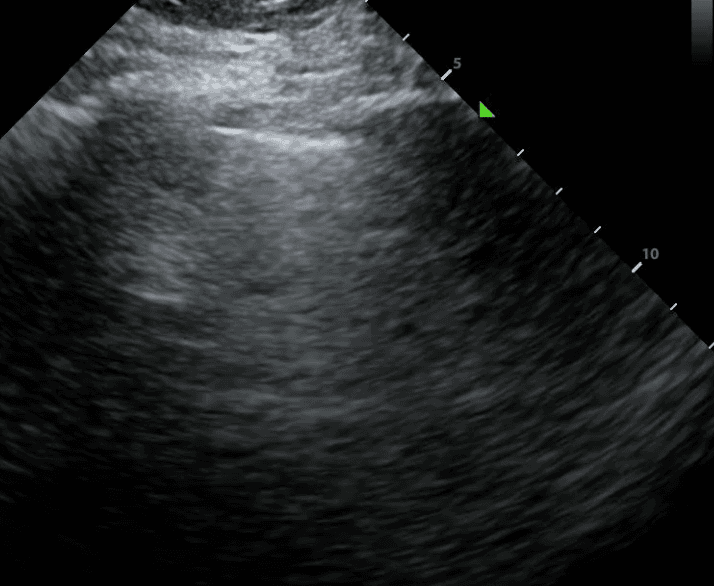
The first step in the 2D assessment of lung sliding is to identify your landmarks. Ribs are discerned at the far left and far right of the above image. They appear as bright (ie. hyperechoic) structures with shadowing below them. In between the rib shadows, you should see the pleural line. This appears as a luminous hyperechoic line. When lung sliding is present, the pleural line has a characteristic shimmering motion that some refer to as the appearance of “ants on a log.”
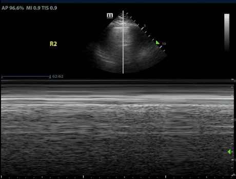
M-mode, or Motion mode, is a type of ultrasound modality that displays motion of a structure through time. In order to detect lung sliding with M-mode, place the M-mode cursor through the pleural line. In the presence of lung sliding, the machine will display an image that is often referred to as the “sea-shore sign” or “waves on a sandy beach.” This stratified appearance is due to the difference in movement between the chest wall versus the pleural line and lung. The motionless chest wall appears as waves while the mobile pleura and lung reflect a sandy beach.

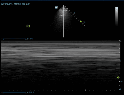
If lung sliding is absent, M-mode will reflect an image of uniform horizontal straight lines known as the “stratosphere sign” or the “barcode sign.” This is because the immobile pleura looks similar to the chest wall.

In a pneumothorax, the air separates the visceral and parietal pleura. Given that the two pleural surfaces are not juxtaposed and moving across one another, there will be an absence of lung sliding. The presence of lung sliding reliably excludes the possibility of a pneumothorax with a negative predictive value of 99.2-100%. However, it is crucial to note that the absence of lung sliding does not necessarily signify a pneumothorax, as conditions other than pneumothorax can also abolish lung sliding.
The differential diagnosis for absent lung sliding can be divided into 3 main categories listed below:
Loss of pleural contact
Fusion of pleural layers
Decreased ventilation
In conclusion, lung sliding is a valuable clinical sign that provides information about normal lung function and aids in the diagnosis of conditions such as pleural effusion and pneumothorax. Its assessment is particularly relevant in emergency and critical care settings where rapid and accurate diagnosis is essential for patient management.
References
Soni, N. J., Arntfield, R., & Kory, P. (2020). Point of care ultrasound. Elsevier. https://shop.elsevier.com/books/point-of-care-ultrasound/soni/978-0-323-54470-2
Radiopaedia, Pneumothorax (ultrasound), accessed 7 December 2023, https://radiopaedia.org/articles/pneumothorax-ultrasound-1.
Bhoil, R., Ahluwalia, A., Chopra, R., Surya, M., & Bhoil, S. (2021). Signs and lines in lung ultrasound. Journal of ultrasonography, 21(86), e225–e233. https://www.ncbi.nlm.nih.gov/pmc/articles/PMC8439137/
Husain, L. F., Hagopian, L., Wayman, D., Baker, W. E., & Carmody, K. A. (2012). Sonographic diagnosis of pneumothorax. Journal of emergencies, trauma, and shock, 5(1), 76–81. https://journals.lww.com/onlinejets/fulltext/2012/05010/sonographic_diagnosis_of_pneumothorax.17.aspx
SHARE
© 2025, DEEP BREATHE, INC